Confocal Microscopy Facility
Research Center
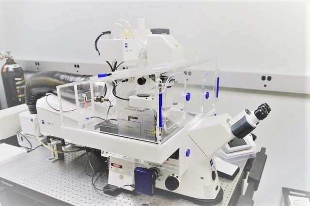
The Center for Dementia Research is equipped with two laser scanning confocal microscopes, an LSM 510 Meta System, and an LSM 880 System, both from Zeiss Microscopy. These systems are used primarily for imaging fixed brain tissue and cell cultures stained with multiple fluorophores. Both systems have heated chambers and CO2 inputs to allow for live imaging of cells in culture with dynamic dyes and can create videos to show changes in real-time. The microscopes have 3 lasers for green red and far-red excitations. The LSM880 can also image blue dyes using a LED and is equipped with the additional capability to make tiled images in a single plane or in z-stacks so that the whole tissue section or cell culture can be reproduced as a 3D rendering. Images can also be analyzed for colocalization between fluorophores or detect relative intensities and sizes of cells and vesicles being studied. CDR confocal microscope analysis has been applied to the human brain and mice modeling Alzheimer’s disease and related disorders (Huntington’s, Parkinson’s, ALS etc.) Cell lines of both human and mouse origins, including fibroblasts from human patients of Familial Alzheimer’s and Down’s syndrome, and primary neurons and blastocysts from mice, are also analyzed in both live imaging and fixed contexts.